The shoulder horn of Dürer´s marvelous Rhinocerus – revealing a 501 year old mystery beast

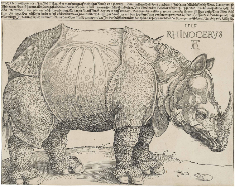
The woodcut of Albrecht Dürer´s „Rhinocerus“ is certainly one of the most iconic animal depictions of the Renaissance. Dating from 1515, a time when books about natural history still usually showed wild mixes of real animals and fully fantastic beings side by side, it is one of the very first depiction of a rhino in European art, and for centuries, one of the most style-defining.

Rhinoceros by Albrecht Dürer, image from Wikipedia

Rhinoceros by Albrecht Dürer, image from Wikipedia

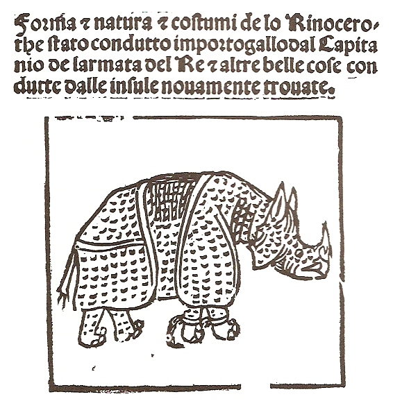
This particular Indian rhino (Rhinoceros unicornis) was a specimen which arrived in 1515 after a long odyssey over sea at Portugal, where it lived in the menagery of Ribeira Palace at Lisbon. The earliest known depiction of the Rhinocerus is a pretty crude drawing by Giovanni Giacomo Penni, a florentine physician who wrote a letter with a poem and a sketch of the rhino, which was published in Rome on 13 July 1515, not even eight weeks after the rhino had arrived at Lisbon.

Rhinocerus at Lisbon by Giovanni Giacomo Penni, 1515. Image from Wikipedia

Not much later,in early 1516, it was shipped again  as a gift from King Manuel I of Portugal to Pope Leo X. Unfortunately the rhino died when the ship on which it was transported shipwrecked off the Italian coast. The carcass of the rhino was later found on the coast of Villefranche.Its skin was preserved and sent back to Lisbon, where it was mounted as a taxidermy specimen. Not much later, the mounted rhino skin made again its way over sea towards Rome, where it finally arrived after its second voyage.

Albrecht Dürer´s woodcut was based on two letters. One was by the merchant Valentin Fernandez, who sent a letter with a descripton of the rhino to a friend at Nürnberg. He had seen the animal himself shortly after its arrival at Lisbon. A second letter from Lisbon, written by an unknown sender, arrived at around the same at Nürnberg and included also sketch of the rhino.
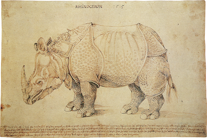
Pen and ink drawing sent to Albrecht Dürer in 1515, collection of the British Museum. Image from Wikipedia

The similarity to the later woodcut is very big, and only a few details, like the fine serration on the backside, were additionally added by Dürer.

Also in 1515, Hans Burgkmair, a German artist from Augsburg, published a woodcut of the Rhinocerus. He had also correspondence with befriended merchants in Lisbon and Nürnberg, but it is not known if he was aware of the same letters and the sketch used by Dürer. His rhinocerus looks much lesser fancy and extravagant as those of Dürer and appears more realistic, but its proportions and features are arguably not even really closer to the life appearance of an Indian rhino than the woodcut by Dürer.
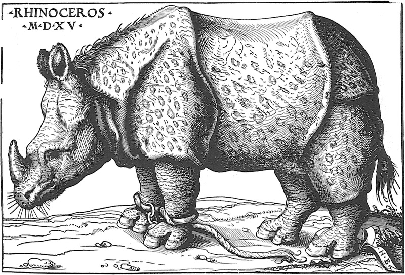
Rhinocerus by Hans Burgkmair, Augsburg 1515. Image from Wikipedia


Dürer´s illustration with the fantastically appearing scales and rivet-like skin pattern, that seems to blend into a crafted yet organic body armour, still amazes with its bizarre beauty. It hast inspired artists for more than half a millenium now, even Salvador Dalí who created a large bronce cast of the rhino. The most unusual feature of the rhino, a tiny twisted horn growing out of its shoulder area, makes it even more appealing to think that Dürer and the unknown illstrator of the foreging pen and ink drawing added a lot of fictional details on the animal for the sake of artistic freedom.

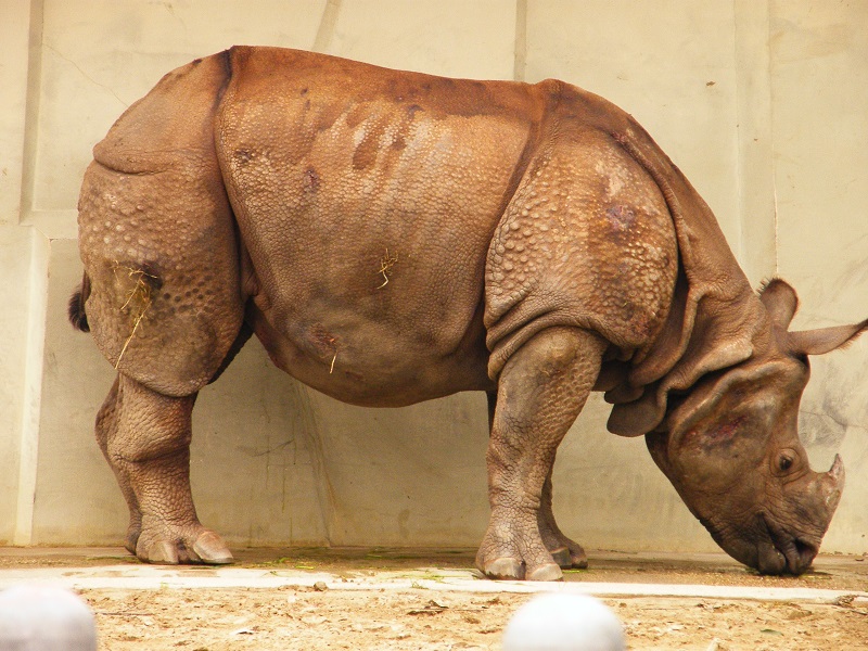
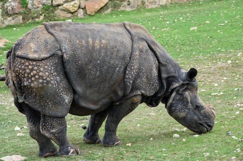
However, those details were probably not even that fictional at all. As weird as the armour-like pattern of the skin looks, it is in fact not that different from the real skin condition of Indian rhinos. Even the gorget-like „neck armour“ is actually not much different from the large neck wrinkles.

Indian Rhino form Wikimedia Commons

Indian Rhino form Wikimedia Commons

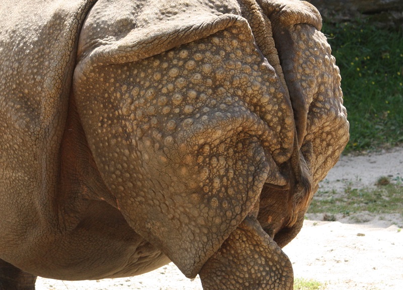
The arrangment of the various skin wrinkles is in the main quite close to the real animal. The rivet-like skin bosses are also, even if somewhat exaggerated, both in size and arrangment mainly consistent. It has been suggested that the unusual skin was a result of the long journey over sea in a small enclosure, which could have resulted into a dermatitis or excessive skin growth, but it is quite hard to say if this is really a convincing explanation, especially since the differences between the depictions and the living animal are not that big at al.

Indian rhino from Wikimedia Commons

Indian rhino from Wikimedia Commons

The rivet-like skin-bosses in detail:

Skin of Indian Rhino from Philadelphia Zoo, modified image from Wikimedia Commons

Skin of Indian Rhino from Philadelphia Zoo, modified image from Wikimedia Commons

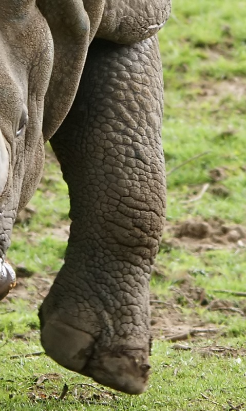
The scale-like skin on the legs of Dürer´s Rhinocoeros, which appears strangley misplaced on a mammal, is also only a comparably subtle modification of the real skin pattern. The gnarly surface of the skin really looks much more like those of a tortoise or other reptile with polygonal scale pattern, and even slightly appears to form overlaying „fish“-scale structures on the upper parts of the legs.

SONY DSC

Indian Rhino leg skin detail, modified image form Wikimedia Commons

So even if the scales on the legs of the Rhinoceros are not fully realistically depicted, they still are not that different from the legs of an actual Indian rhino, even if Dürer and the original illustrator clearly exaggerated and altered the amount and structure of the rhino´s skin to some degree.

For more than a century Dürer´s woodcut was by far the most influential source for depictions of rhinos and was more or less accurately copied for countless times. Even in the 18th century the legacy of the Dürer rhino was found in artistic depiction.

Alessandro de’ Medici even included the rhino, which was obviously highly influenced by Dürer´s woodcut, into his coat of arms.
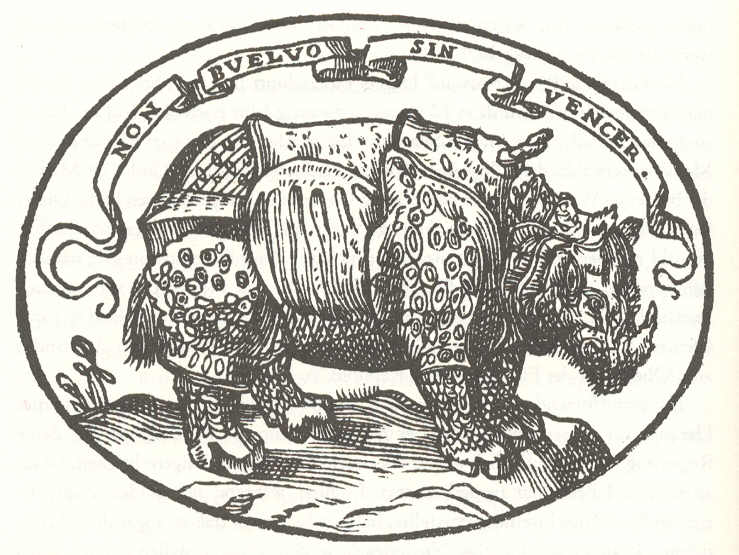
Coat of arms of Alessandro de’ Medici. Image from Wikipedia

But what about the most intriguing feature of Dürer´s rhino, the spiraling horn growing out of its shoulder? Ancient artists and scholars who based their description mainly on the few already existing artworks and reports assumed that this horn was really existing. Some even speculated that it was used in intraspecific combats. Some of those depictions show the dorsal horn in an even more twisted and bigger way than the original drawing and woodcut, and there is at least one depiction which shows also a second smaller dorsal horn. The question remains, was it simply a fully imaginary rendition of the unknown illustrator who drew the reference for Dürer´s woodcut at Lisbon?

Well, probably not, at least it was not a fully fictional reinvention. Small horn-like structures are actually known from from white rhinos (Ceratotherium simum), as documented by Bernhard Grzimek and later Karl Shuker (Extraordinairy Animals Revisited, 2007). Based on reports of white rhinos with unusual horns growing on their bodies, Bernhard Grzimek already suggested decades ago that there was possibly a connection with the shoulder horn of Dürer´s Rhinoceros (Thanks to Karl Shuker, who provided me this information). Here is for example a white rhino from Lake Nakuru National Park, which shows three small ceratinous skin growths in its neck area:
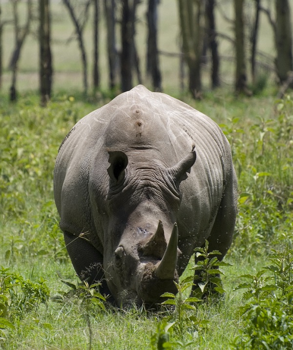
White rhino, photo from Wikimedia Commons.

White rhino, photo from Wikimedia Commons.

Detail of the shoulder area:
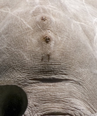
White_Rhinoceros_in_Lake_Nakuru_National_Park,_February_2007 detail

Similar hyperceratoses in the shoulder area also occur in Indian rhinos.
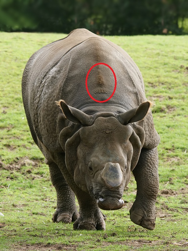
Indian rhino with small hyperceratosis in shoulder area. Modified photo from Wikimedia Commons

Indian rhino with small hyperceratosis in shoulder area. Modified photo from Wikimedia Commons

Another detail photo:
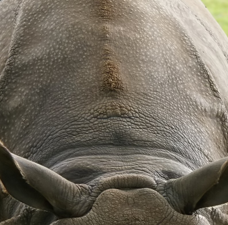
SONY DSC

Indian rhino with hyperceratosis in shoulder area

This small areas of excessive skin growths are however still far away from something like a horn, not to mention a twisted narwhale-tusk like horn. But there are even much more extreme cases. When I was many years ago at the zoological collection at Hamburg, I noticed a taxidermied Indian rhino in the collection which had a strange outgrowth in its upper neck area. A row of blunt ceratinous knobs and spikes, from which one was even forming a small horn-like structure.
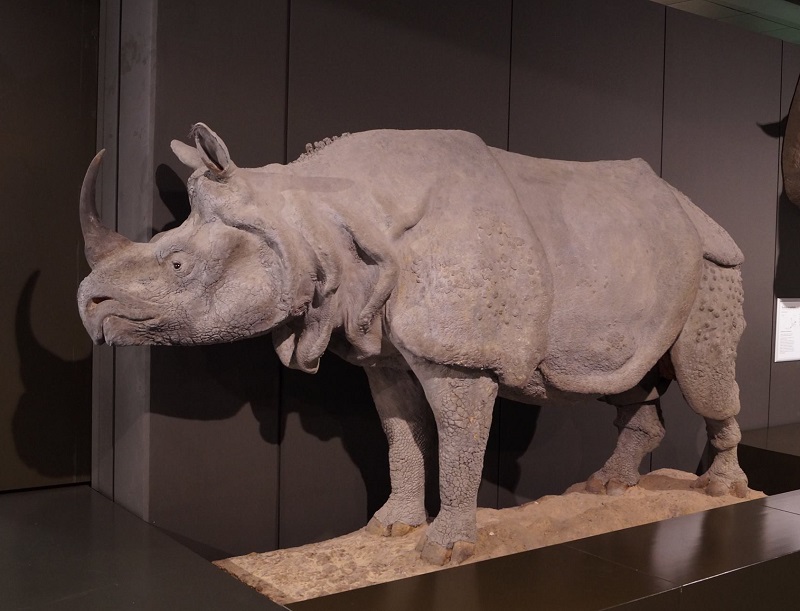
Indian Rhino, Zoological Collection Hamburg. Photo by Sven Sachs, Naturkundemuseum Bielefeld

At that time I sadly took no photo of the whole specimen, but luckily my good friend Sven Sachs had one from a recent visit of the museum and gave me kindly permission to use it for my blog. Here is a photo I took of the head and neck area:
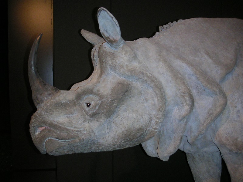
Indian rhino with shouldern horns, Zoological Collection Hamburg. Photo Markus Bühler

A closer look:
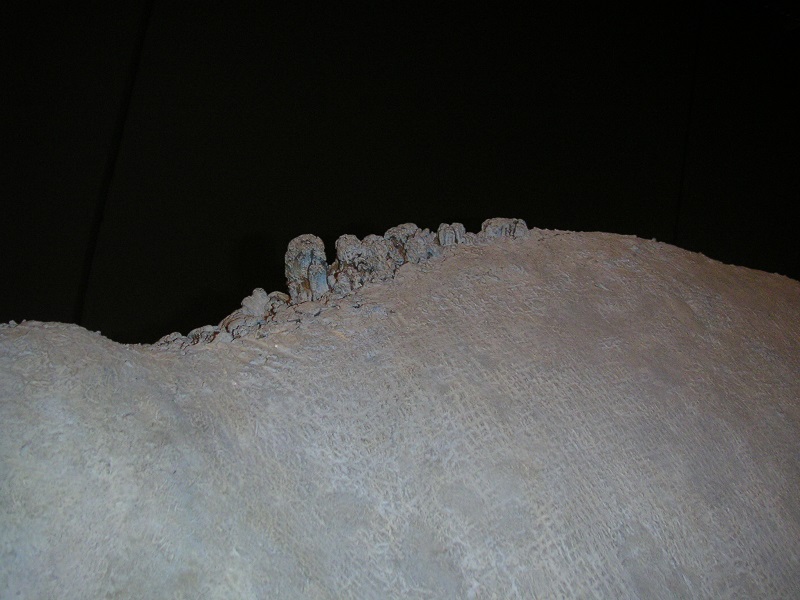
MINOLTA DIGITAL CAMERA

This photo shows the small blunt „horn“

A somewhat modified close-up which shows the surface structure. The „horns“ seems comparably massive and there is also apparantly some wear on their surface.
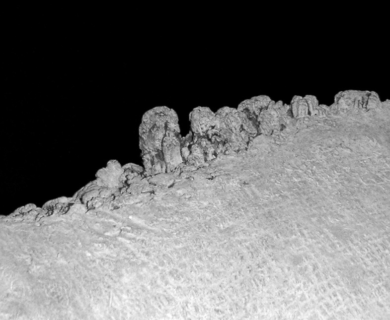
Detail of ceratinous shoulder horn and humps

Another photo of the rhino which I took in a more frontal view:
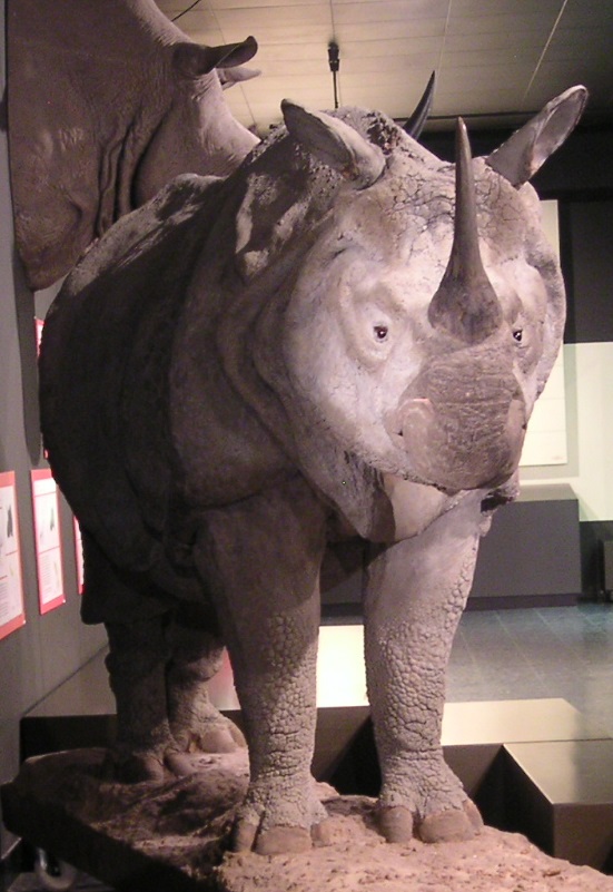
Indian rhino with shoulder horns, Zoological Collection Hamburg, Photo Markus Bühler

Indian rhino with shoulder horns, Zoological Collection Hamburg, Photo Markus Bühler

I can not say what causes this hyperceratosis, if it occurs mainly in very old specimens or if it is even a pathological condition, some sort of skin reaction on external irritations, or if similar growth processes like those of the cranial horns are on work in misplaced areas. A histological examination of such a skin or reports of captive specimens could give probably more answers. The presence of nearly perfectly formed neck- or shoulder horns in some white rhinos indicates however that there actually is a connection of some sort with the cranial horns.

Another case of three comparably large neck-horns in a white rhino from Duisburg Zoo can be seen here. It was a nearly 50 years old female named Nongoma which died in 2014.

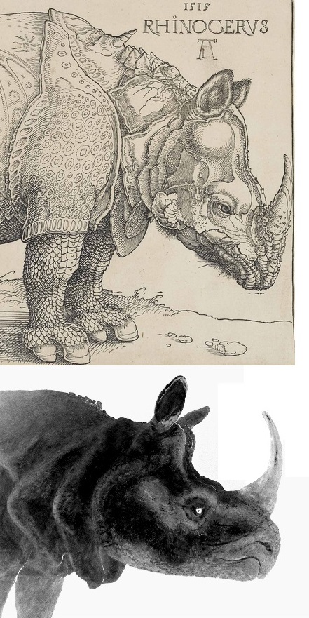
A comparison of Dürer´s woodcut with a graphically modified photo the taxidermy specimen from Hamburg. I think it is also noteworthy that there are in front of the spiraled horn also some other, smaller outgrowths on the neck, which are in line with the multiple areas of hyperceratosis found in some of the rhinos shown above.
Dürernashorn Vergleich 2

The twisted horn of the Rhinocerus is clearly a product of artistic freedom, but the horn-and boss-like structures, which really are present in some individual rhinos, show that it was not just a fully fictional decorative element. It would have been a quite extreme case of coincidence if somebody just invented a shoulder horn for rhinos without any knowledge that similar outgrowths really occur in exactly that area. Perhaps there were already older reports from Asia or even Africa which mentioned those shoulder horns, but there is a chance that the rhino at Lisabon was really one of the rare specimens which had such a condition.

(Update): I was also just informed by Connor Lachmanec about a white rhino with a neck or shoulder horn in Vancouver Zoo. This rhino named Charlie has a pretty enormous horn growing of of its neck, which is not just an amorphous growth or callus but a nearly perfectly formed horn. It seems there were possibly once even two other horns which are now broken off. See the amazing photos of Charlie here, here and here.

The simple sketch by Giovanni Giacomo Penni shows nothing that would indicate such a thing. The more detailed woodcut by Hans Burgkmair shows also no horn-like structures over the shoulder or neck, but interestingly he added a mane of bristles on the top of the neck, a feature not found in Indian rhinos. Was it a reinvented detail, perhaps because Burgkmair or his correspondents assumed this animal was somehow similar to a wild boar? Or was it possibly an interpretation of really existing gnarly growth on the neck of this rhino?

We will never know for sure of course. But after this detailed look at the most famous rhino in art history, the Rhinocerus of Albrecht Dürer appears no more that fantastic and made-up as usually assumed, and even some of its more bizarre features like the shoulder horn have quite probably a real origin. So more than 500 years after Albrecht Dürer published the famous woodcut, his marvelous beast has finally redeemed itself to some degree from its overly fantastically appearing depiction.
 

Veröffentlicht unter Blogposts in English, Kryptozoologie, Populäre Irrtümer, Säugetiere | 6 Kommentare

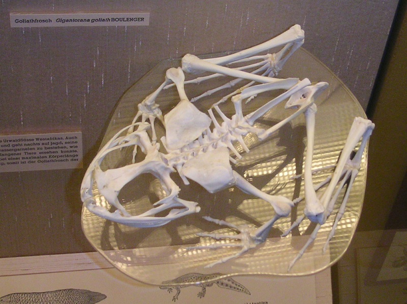
Photo of the day: Goliath frog skeleton

Today´s photo of the day shows a skeleton of a goliath frog (Conraua goliath) from the zoological collection at Heidelberg:

Goliath frog skeleton (Conraua goliath), zoological collection Heidelberg.

Goliath frog skeleton (Conraua goliath), zoological collection Heidelberg.

Veröffentlicht unter Amphibien, Anatomie, Bild des Tages | Schreib einen Kommentar

Anglerfish on ice

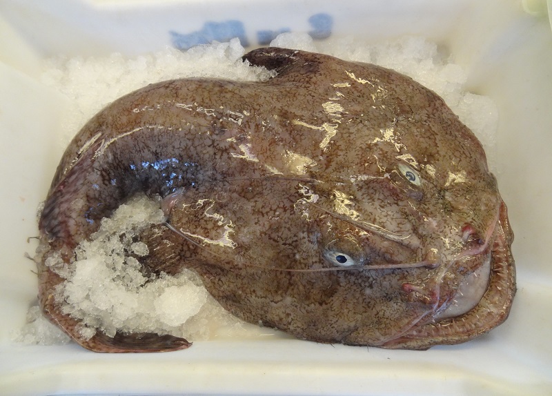
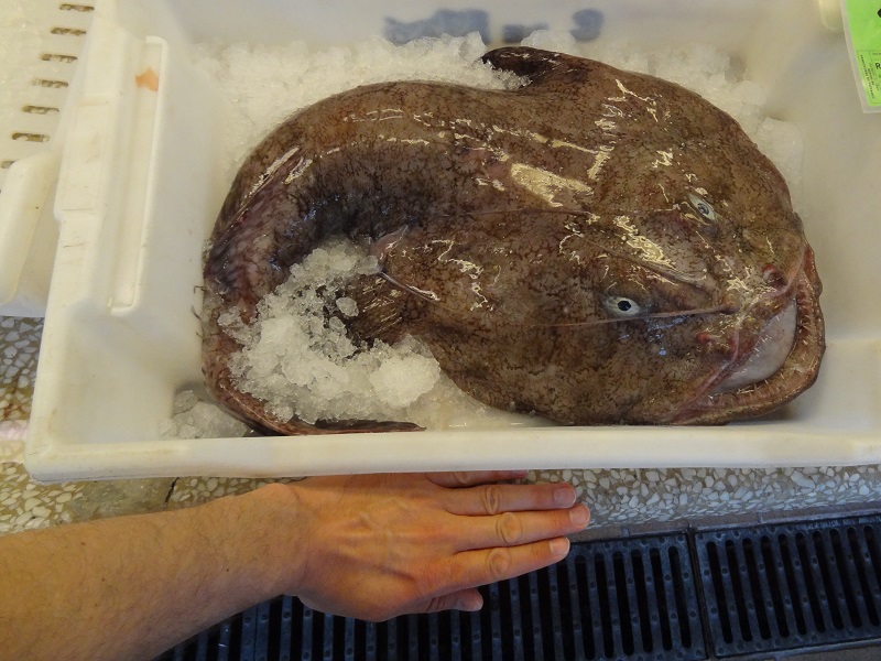
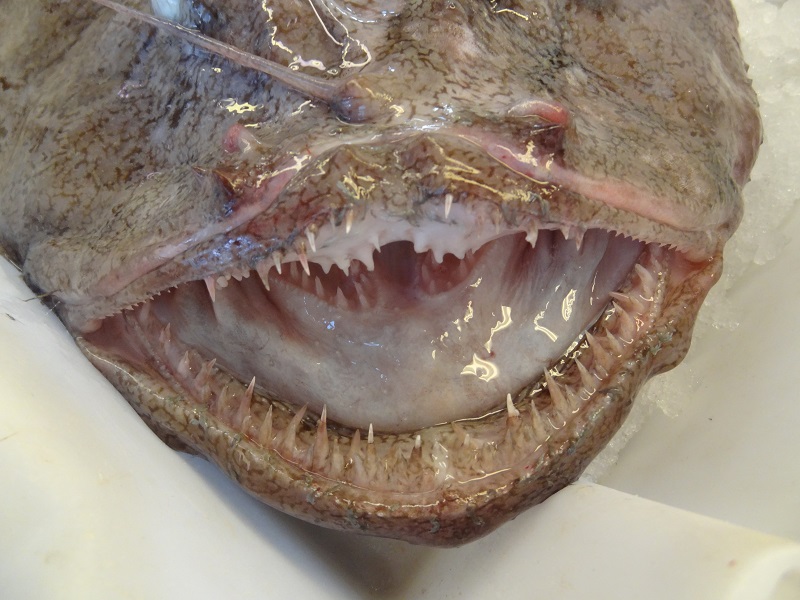
Besides strange flatfish, the fish market at Mahón had also a lot of other interesting species on display, like several monkfish (Piscatorius lophius), including one of the very largest specimens I have ever seen. I covered monkfish already some times before on the blog, for example for their occasional predation on birds, highly modified skeletons or general weirdness.

Monkfish (Lophius piscatorius) on fish market at Mahón

Monkfish (Lophius piscatorius) on fish market at Mahón

This specimen was still far away from the upper sizes which this species can reach (more than 1,5 m in exceptional cases), but even the sheer sizes if its head was already staggering.

Lophius piscatorius (2)

A close photo of the bear trap alike jaws which shows also the large set of teeth on the tongue bones.

Lophius piscatorius (3)

Monkfish jaws

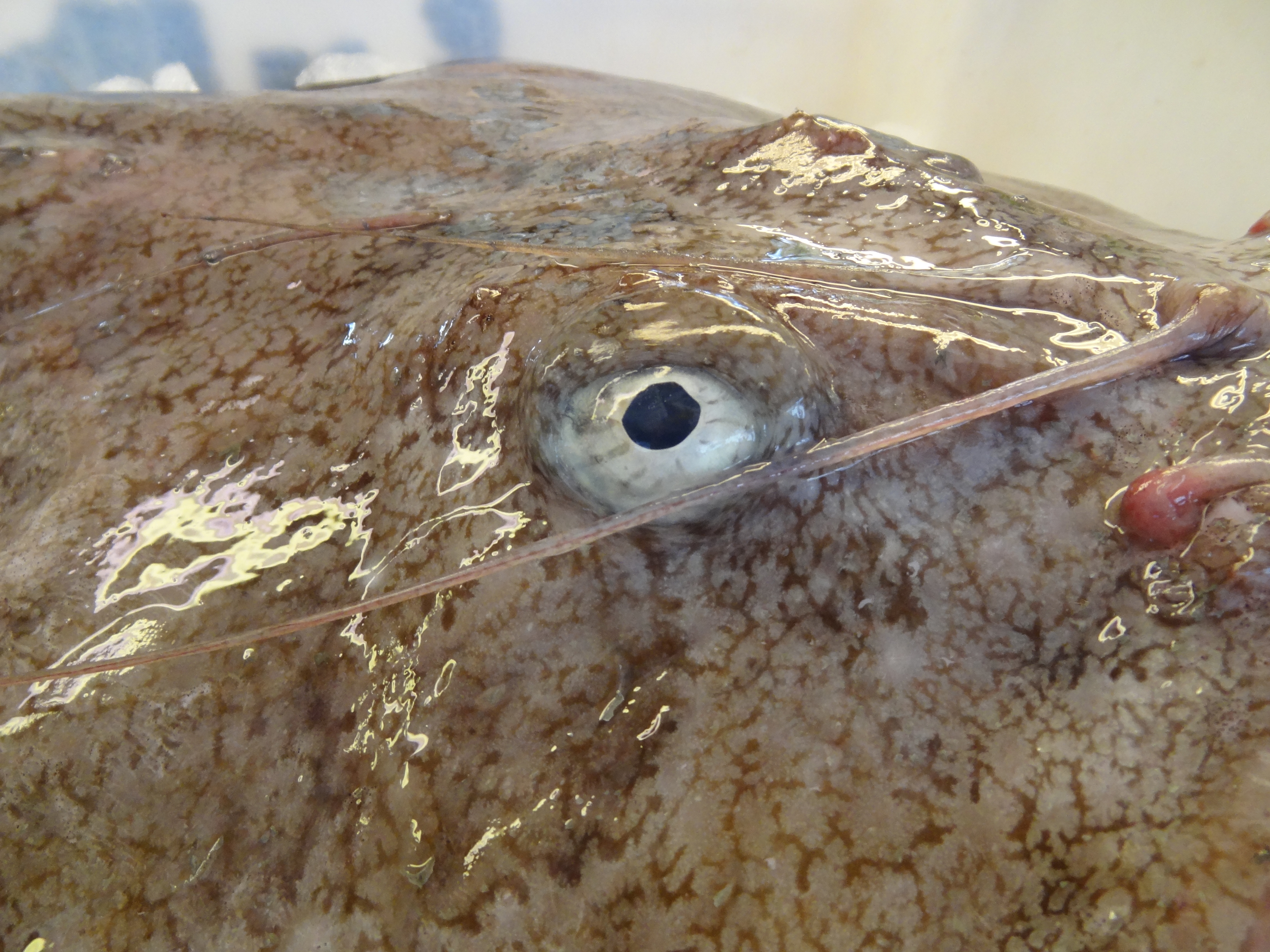
Detail photo of one of the eyes.

Lophius piscatorius (5)

Monkfish eye

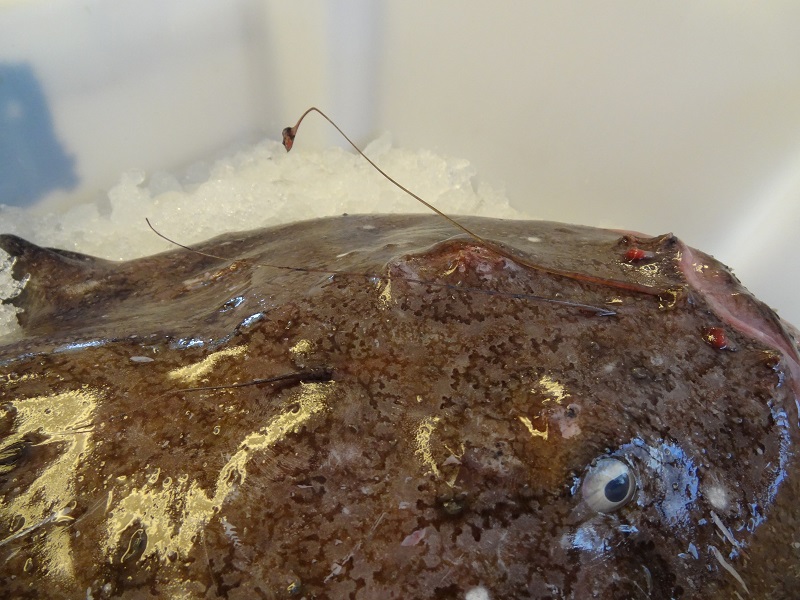
A second, somewhat smaller specimen which shows the rod or illicium with the skin appendix acting as bait.

Lophius piscatorius (6)

Monkfish illicium

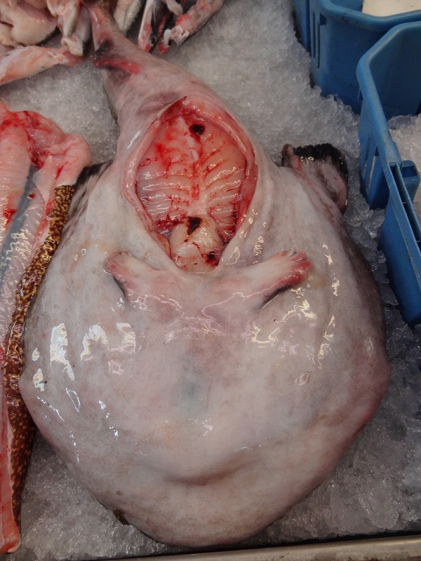
Lophius piscatorius is easily distinguishable from the quite similarly looking black-bellied angler (Lophius budegassa) when its abdominal cavity is opened. In L. piscatorius the membrane inside the cavity is whiteish, and blackish in L. budegassa.

Monkfish with opened belly and visible ventral fins

Monkfish with opened belly and visible ventral fins

You can also see the two fleshy hand-like ventral fins, which are used by the monkfish to move on the bottom.


Veröffentlicht unter Blogposts in English, Fische | Schreib einen Kommentar

Photo of the day: Lepidorhombus boscii

Fish markets can be a great opportunity to see animals you will hardly ever see in any zoo aquarium or in TV. Alcohol specimens in museum collection are often already far away from their original life appearance, because the tissue shrinks to some degree, they lose most of their colouration, transclucience and other features of the living animal you can´t even fully reproduce in a really good painted taxidermy specimen or life-like model. Seeing an animal in the flesh, and not just its preserved and modified body, can teach you much more about its physiology than a faded specimen in a jar. So I was quite eager to take photos when I was recently visiting a fish market at Mahón, the capital of Menorca.

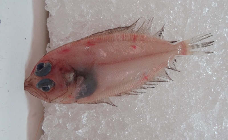
One of the very first fish I disovered on display was a quite weird-looking flatfish of around 15-20 cm, with a nearly colourless and partially translucient body, huge bulging eyes and long jaws. It seems to be Lepidorhombus boscii, a close relative of the megrim or whiff (Lepidorhombus whiffiagonis). Sadly the dorsal and anal fin were quite damaged, but it seems there were originally two dark spots in the area close to the caudal fin, what would indicate that this was really L. boscii.

Lepidorhombus boscii

Lepidorhombus boscii

Flatfish are already bizarre per se, after all, they have some of the most radical anatomical modifications among all chordates at least. Keep this in mind if you´re eating the next time a sole or a halibut, this animals have evolved into something Pablo Picasso could have invented in a fever-dream.

Veröffentlicht unter Bild des Tages, Blogposts in English, Fische | Schreib einen Kommentar

Photo of the day: Ghastly tenrec taxidermy

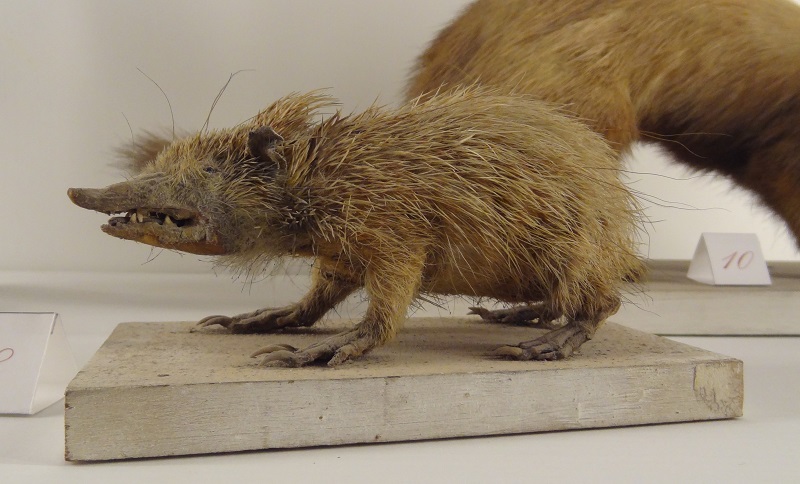
For some reason tenrecs are especially prone to become extremely ghastly taxidermy specimens, at least if you look at the older ones. I just planned to cover them on occasion on the blog. So here is a specimens I saw last week at the Museu Diocesà de Menorca at Cituadella, within an old zoological collection:

Tenrec ecaudatus taxidermy specimen at the Museu Diocesà de Menorca, Cituadella

Tenrec ecaudatus taxidermy specimen at the Museu Diocesà de Menorca, Cituadella

Veröffentlicht unter Bild des Tages, Blogposts in English, Ghastly tenrec taxidermy, Säugetiere | Schreib einen Kommentar

Of sea monsters and cetacean weirdness – here´s the skeleton of a long-finned pilot whale

Based on the recent discovery of an alleged „monster“ on a British beach, I decided to post a photo of a long-finned pilot whale´s skeleton from the zoological museum Hamburg. First of all, because that´s exactly what this purported monster really was, a highly decomposed and already partially burned skeleton of a long-finned pilot whale (Globicephala melaena). It is fully beyond me why so many people somehow forget that a lot of animals have bones at the inside, and if the overlaying soft tissue decomposes, mummifies or gets eaten away by scavengers those „bones“ can become visible and the whole thing looks different from its life-appearance.

Decomposed whale carcasses have already fooled countless people, especially when the bare bones of the caudal vertebrae were visible, what appeared to many like a crocodile-like tail. Furthermore the head and skull shape of many ododontocetes look very different, and even quite bulbous-headed species like belugas, orcas or pilot whales easily appear reptilian to people not familiar with their cranial anatomy. Even more so if the lips and the cheek tissue are already gone, and a surprisingly number of surprisingly big teeth is suddenly visible. But that´s not the only reason why I post this photo of a long-finned pilot whale´s skeleton.
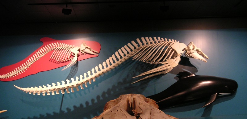
It´s also because this skeleton shows well just how freaky Globicephala melaena really is, especially when compared with its close relative, the short-finned pilot whale Globicephala macrorhynchus (visible on the left).

Skeleton of long-finned pilot whale at the zoological museum Hamburg

G. macrorhynchus has a quite conservative body shape, which doesn´t differ that much from most other Delphinidae. But G. melaena has not only an extremely elongated body which looks almost a bit like something on the way to evolve into a Basilosaurus, it has also this enormously elongated pectoral fins with nearly wing-like finger bones.

If we would know it only from its fossils, it would probably be counted as a real weirdo, in line with Basilosaurus, Odobenocetops or Eurhinodelphis. However, it´s not, perhaps just because it´s a too familiar whale, which has long been known to many European coastal nations. Sadly we easily tend to overlook the unusual and special in animals which are apparantly too familiar or too normal.

Veröffentlicht unter Anatomie, Blogposts in English, Kryptozoologie, Wale | 2 Kommentare

Starting into the herpetology season part III: On the track of green lizards

Green lizards are not only the largest but also surely the most flamboyant lacertids of central Europe. Besides this, they are also the rarest kind of lizards here around, with only a few handful of often quite local populations. Fortunately, one of those local populations is living nearly in sight from where I live. This population had also a somewhat cryptic status and was next to unknown for a very long time. So far I had only seen one single specimen within three decades, about which I wrote here.

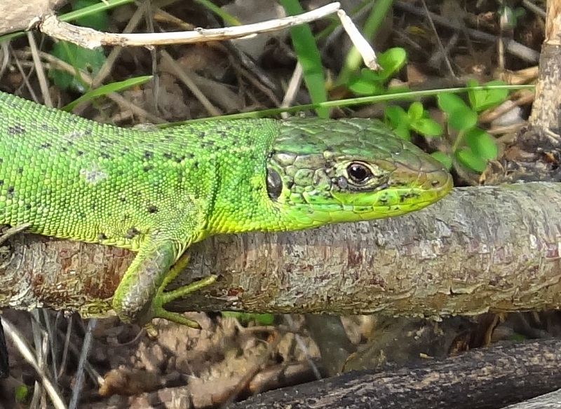
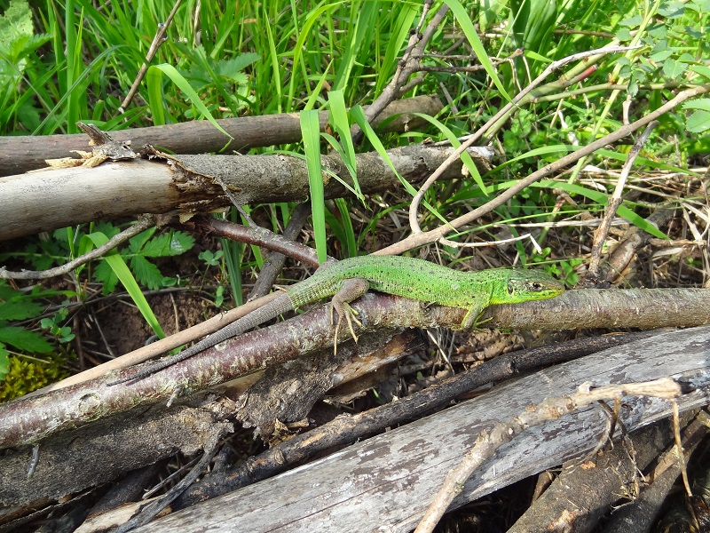
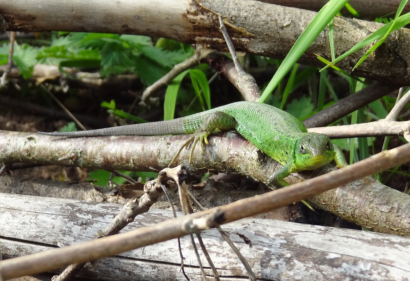
But three days ago I made once again a long walk in this area, and was highly delighted to discover two more specimens. One of them was a subadult from which I could take some really close photos.

Smaragdeidechse 2

As you can see, it had a regenerated tail.

Smaragdeidechse 1

A view from another perspective:

Smaragdeidechse 4

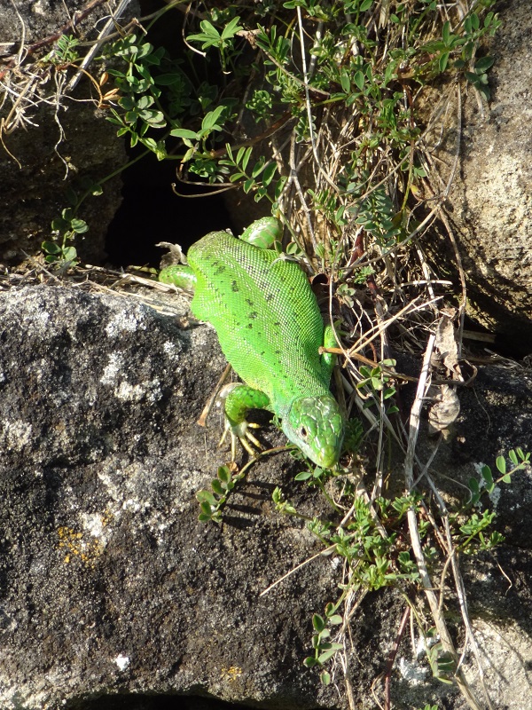
The second specimen which was basking on a wall was well bigger, and as I could later see on the photos, had just shed its skin.
Smaragdeidechse 3

I think it´s still not fully known to which of the green lizard species this population belongs to, as the Western green lizard (Lacerta bilineata) and the Eastern green lizard (Lacerta viridis) are looking extremely similar. Furthermore, both species occur locally in Germany.
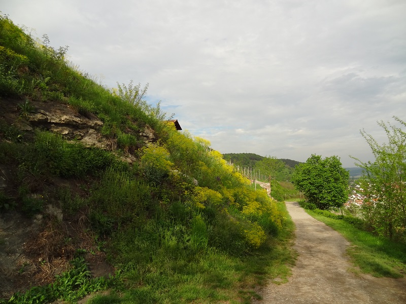
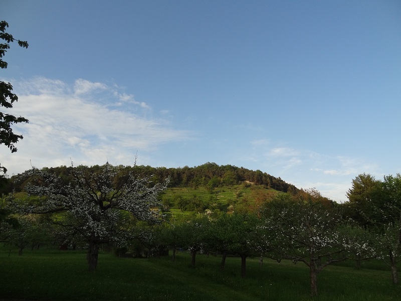
This particular area has a very warm microclimate and houses a lot of rare animals and especially plants.

DSC04812

Furthermore the landscape is quite diverse, with vineyards and meadows, orchards, shrubs and also forested areas.

DSC04947

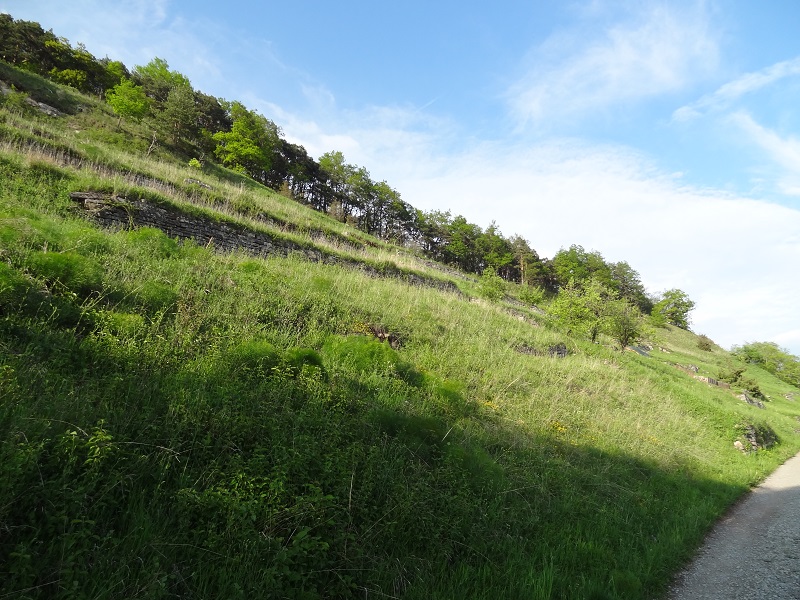
Originally most of this slope was covered with vineyards, but today only dry walls remain on big areas, making them perfect habitats for many species.

DSC04924

One of the wonderful flowers like this military orchid (Orchis militaris) which are growing there:

DSC04929

If you take a very close look on them, they look like tiny elves cut from paper.

DSC049292

You can see more photos of green lizards which I took in the Kaiserstuhl area in another blogpost.

Veröffentlicht unter Naturbeobachtungen, Reptilien | 2 Kommentare

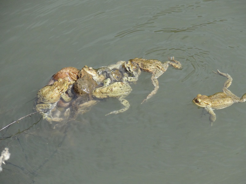
Starting into the herpetology season part II: Orgiastic toad balls

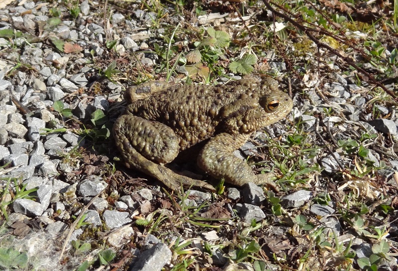
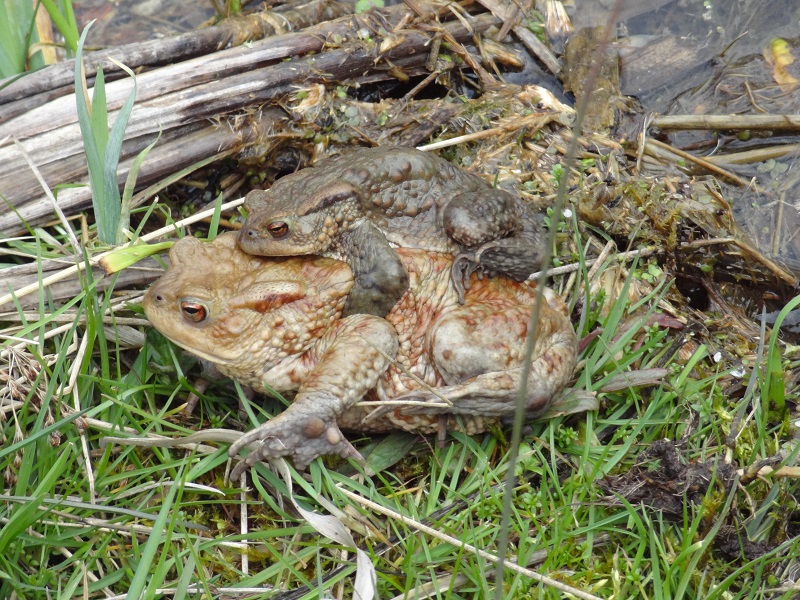
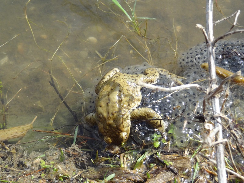
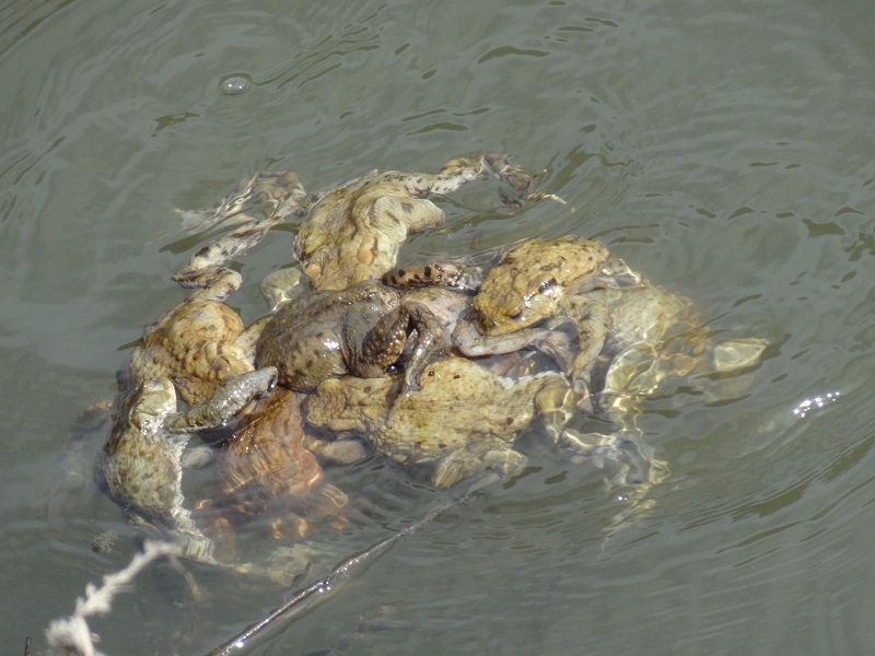
Mass gatherings of mating common toads (Bufo bufo) are probably among the most spectacular herpetological phenomens you can see among European amphibians. It is really an incredibly sight to see dozens or even hundreds of this usually quite solitary anurans gathered together. It is not only interesting to see such a large number of individuals with all their range of intraspecific variation, but also to see a lot of fascinating, weird and sometimes even quite obscene behavior.

DSC04357

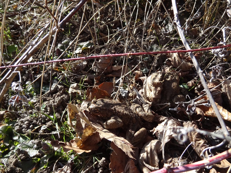
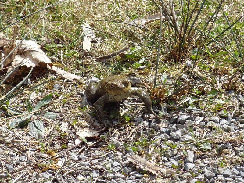
At the end of March I visited a pond which is every spring visited by mating toads. I found only a few specimens in the water, some toads were even still walking or sitting around on land set back from the pond and apparantly on their last steps of their journey to the mating ground.

DSC04362

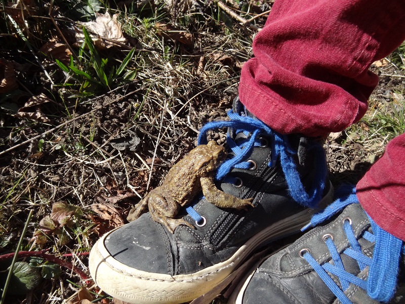
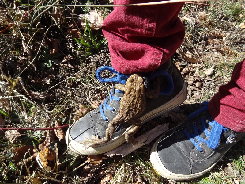
One male toad jumped on the shoe of a young boy who visited the pond with his grandparents. I don´t think he knew what the toad was going to do when it clinged to his foot.

DSC04363

It tried to perform the amplexus, a mating behavior in which the males grab the females with their arms and cling on their backs.

DSC04364

Some of those male toads have a quite extreme sexual urge, and are quick to try to mate with just anything, including other amphibians or even inanimate objects.

When I came only a few days later again on the pond, the weather was now already much warmer, it was just full of toads, sitting and swimming around, and of course – mating.

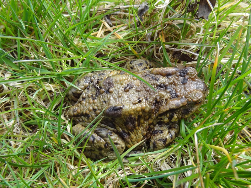
Here is a photo of a large female which was sitting in the gras, surprisingly still without a male.

DSC04383

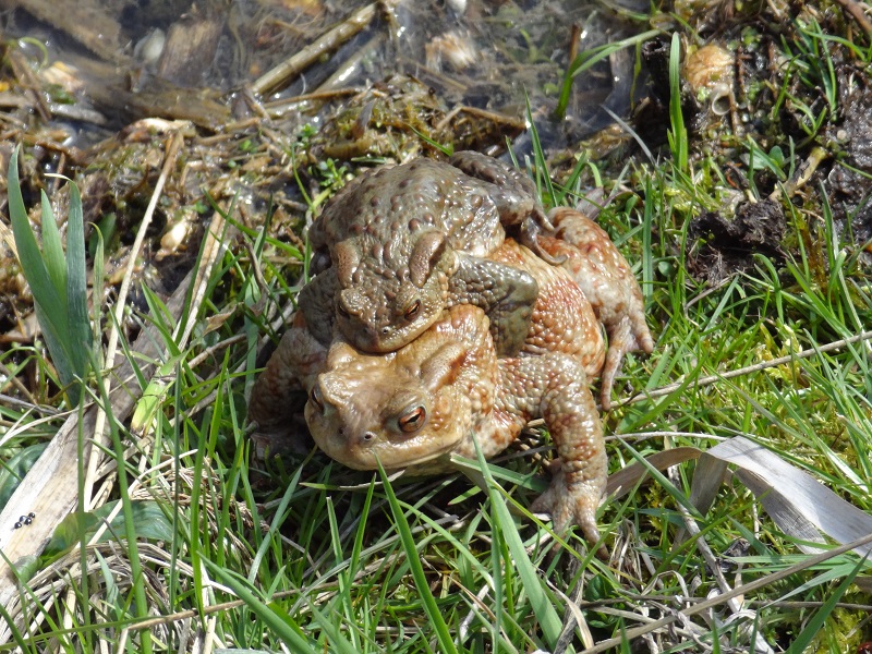
Other females had already found a fitting male, like this happy couple shows:

DSC04427

This photo shows also quite well the strong sexual dimorphism:

DSC04487

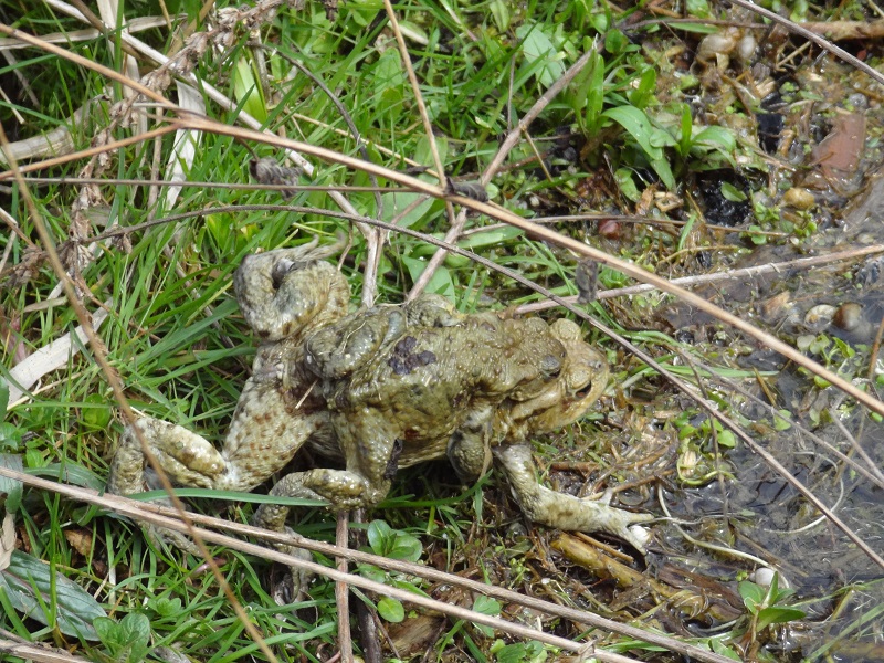
It also looks really strange when the females and males are walking together in this posture.

DSC04514

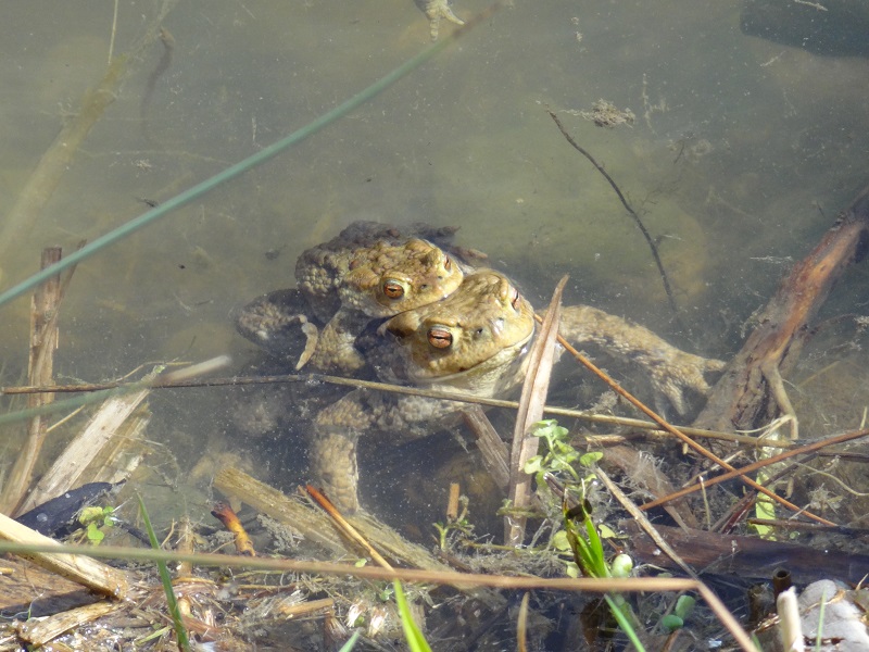
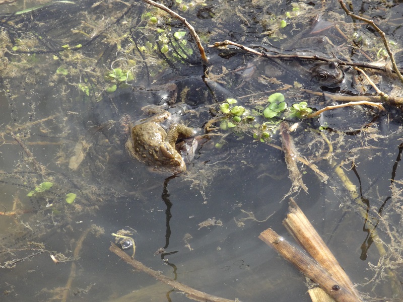
Other pairs were already together in the water.

DSC04404

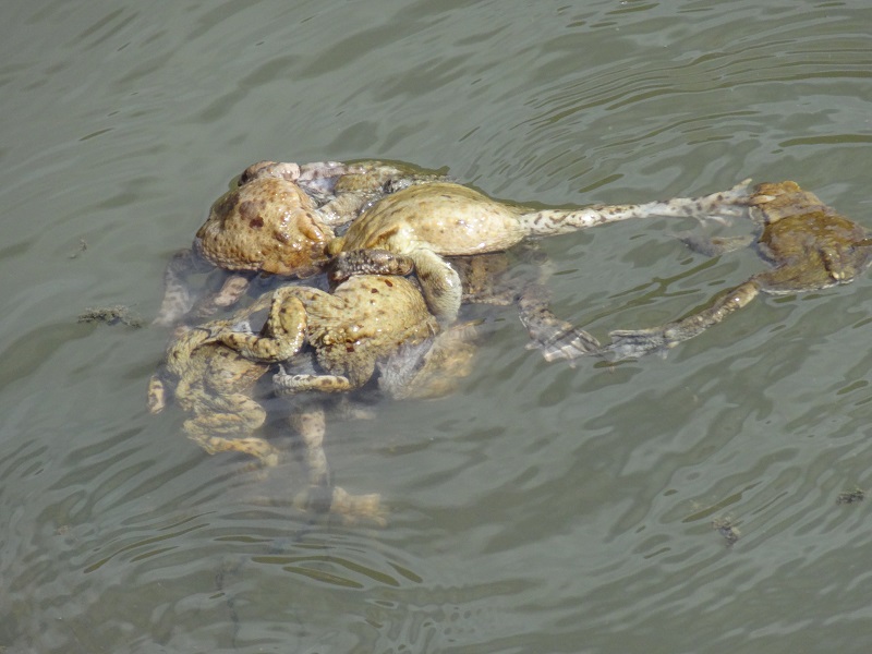
Other females had lesser luck and were literally sieged by horny toad males.

DSC04458

The females in those balls were no more visible, and it can happen on occasion that they drown under such masses of wannabe-lovers.

DSC04455

There is even a case reported in which an adult male common moorhen (Gallinula chloropus) nearly drowned because several male toads clinged on its head and neck.

DSC04452

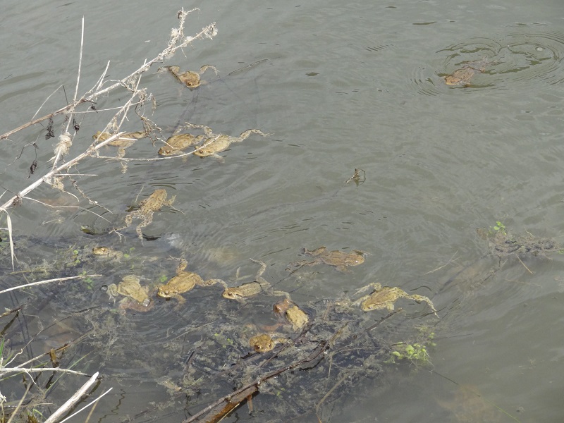
Another photo showing the large numbers of male toads in some areas of the pond.

DSC04490

This obviously quite frustrated male continuously tried to copulate with a clump of moor frog (Rana arvalis) spawn.

DSC04477

I could also observe a toad male which tried to jump on a moor frog, but apparantly quickly realized his error. You can also see a lot of egg strings around the toad and the two moor frogs.

DSC04414

Another opportunistic toad male jumped out of the water up to me and chased me for several metres. That´s really a very weird feeling if a tiny toads is chasing you. Somewhat later another toad also suddenly chased me, or perhaps more probably, my shoes, which must have appeared to him like a potential female toad.

DSC04483

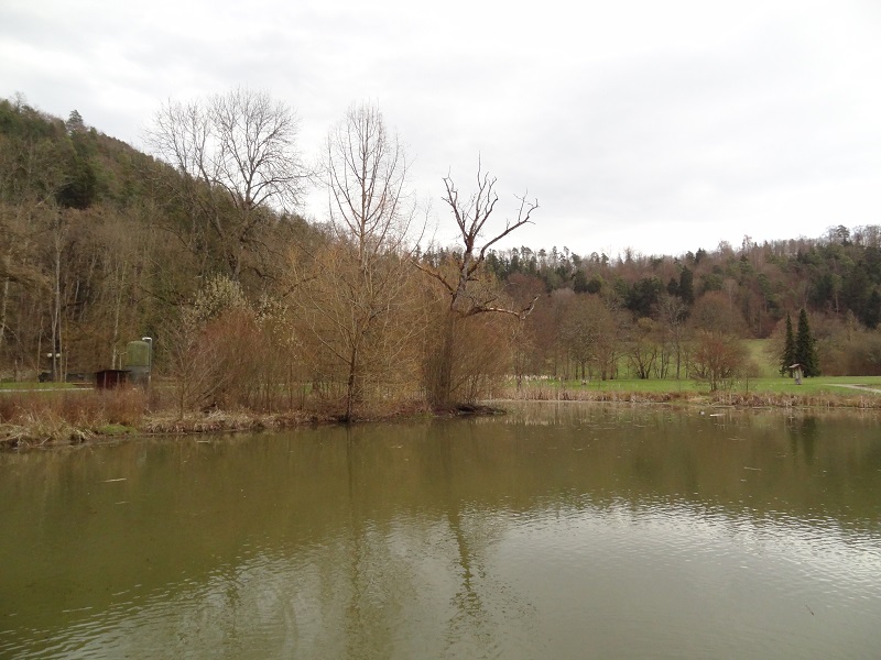
Here is a photo of the pond, which is located in a small park surrounded by woods.

DSC04499

Meanwhile I have already seen the first tadpoles, and within a short time, the whole pond will be full of them.

Veröffentlicht unter Amphibien, Naturbeobachtungen | 3 Kommentare

Starting into the herpetology season part I: Newts are coming

After the last entry spontaneously disappeared due to a server problem (together with the draft of the next part of the series), I make a somewhat shortened second version to restore the first part again.

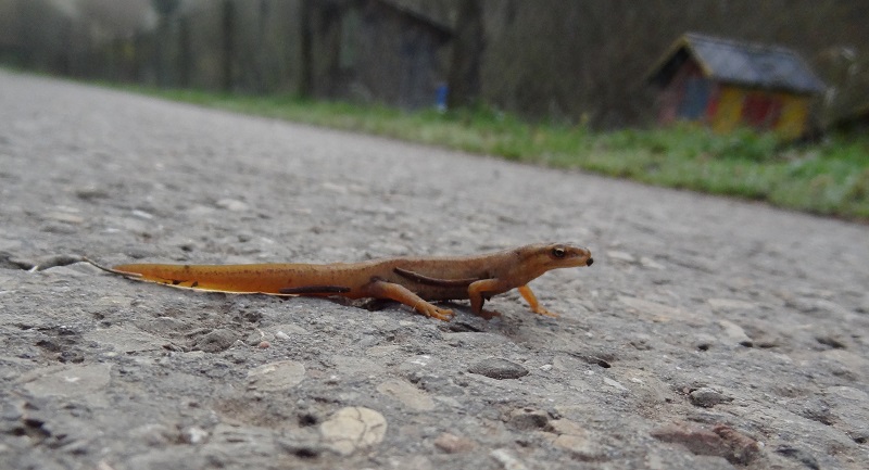
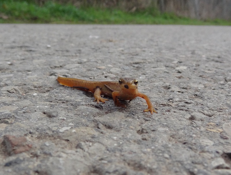
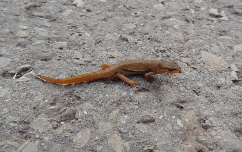
So here are some photos I took two days ago during a walk, when I encountered a common newt (Lissotriton vulgaris) sitting on an asphalt field road. It was probably on its way to a pond or ditch. It was one of the first amphibian species I´ve seen this year, after I discovered the first alpine newts, common toads and moor frogs only a few days before.

Common newt Lissotriton vulgaris (2)

Common newts are besides alpine newts (Ichthyosaura alpestris, a pretty awesome name BTW) by far the most common caudates here around.

Common newt Lissotriton vulgaris (3)

In their terrestrial form common newts are sometimes mistaken for lizards by people, because their dry and finely granulated skin doesn´t appear very amphibian. Furthermore their tails have not the large skin appendiges which form the fins and crests which develop in the aquatic form during the mating season.

Common newt Lissotriton vulgaris (1)

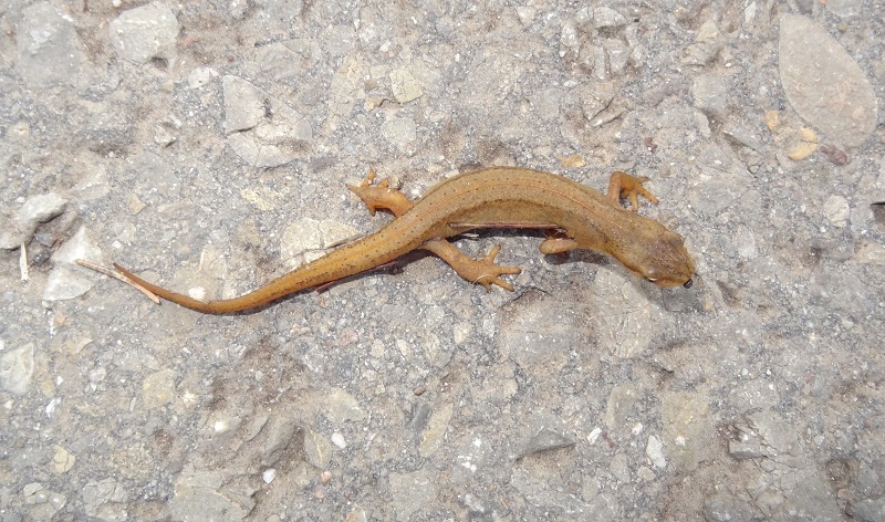
Another photo showing the specimen in dorsal view:

Common newt Lissotriton vulgaris (4)

After I took the photos I carefully placed it in the nearby gras, as this road is comparably strongly frequented by cyclists, joggers and also cars, what sadly sometimes results in a lot of roadkilled amphibians at early morning, especially of fire salamanders.

Veröffentlicht unter Amphibien, Naturbeobachtungen | Schreib einen Kommentar

Under the hermit crab´s shell

Hermit crabs are freaky and cool for many reasons. They live inside the external skeletons of dead snails to cover their misshapen abdomens, which they even sometimes plant with living anemones, they have some of the most extensive body assymetries of all crustaceans, some of them are among the most succesful terrestrial decapops of the world while their largest member, the coconut crab Birgus latro is by far the largest terrestrial arthropod since the Carboniferus.

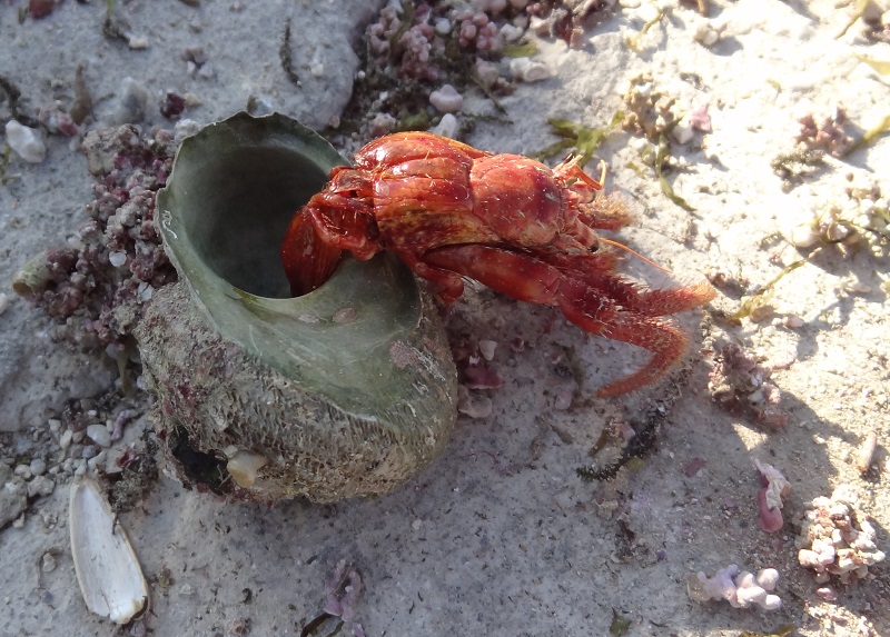
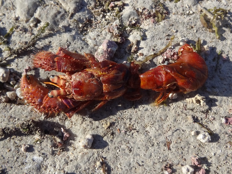
When I was two years ago at Crete I had the chance to take a closer look at two dead hermit crabs from some fishermen´s bycatch.

DSC06163

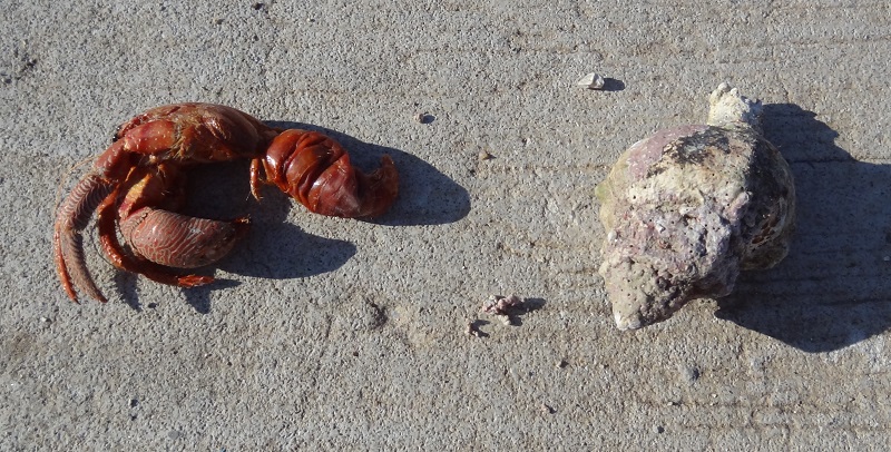
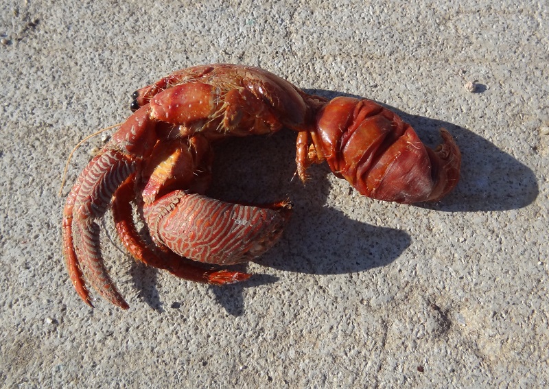
You can usually only see the front part of the body, but in this dead individuals I could easily separate the hermit crabs from their shells to take a look at their abdomens.

DSC06164

They had been lying there already since some time in the sun, so the very soft abdomens were already somewhat shrunken.

DSC06165

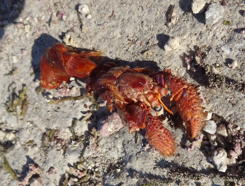
A look at the other specimen:

DSC06162

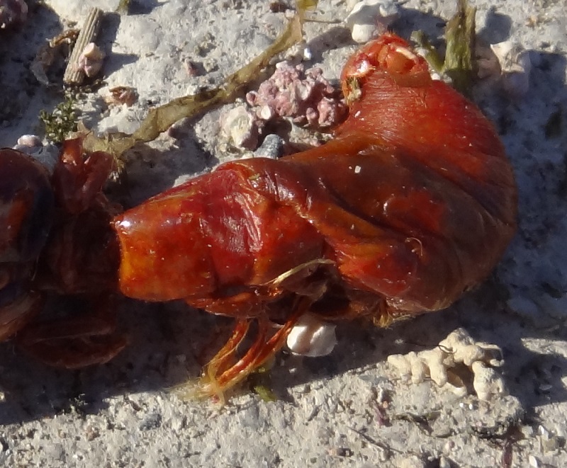
On the left side of the abdomen you can also see the highly modified pleopods used to hold the abdomen inside the shells.

DSC06160

Close-up of the abdomen:

DSC06160 2

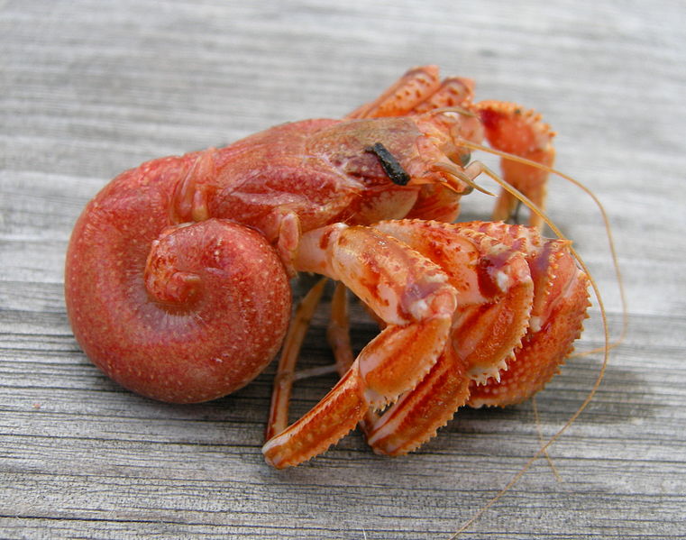
For comparison a photo of a quite fresh Pagurus bernhardus from Wikipedia, with the abdomen still in full shape:

Pagurus_bernhardus

Pagurus bernhardus. Source: Wikipedia

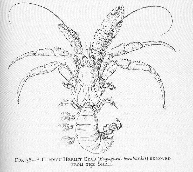
Here´s also for the better understanding of the anatomy a schematic depiction of a Hermit crab without shell:

FMIB_46415_Common_Hermit_Crab_(Eupagurus_bernhardus)_removed_from_the_shell

Common Hermit Crab (Eupagurus bernhardus). Source: Wikimedia Commons)


Veröffentlicht unter Anatomie, Arthropoden | Ein Kommentar